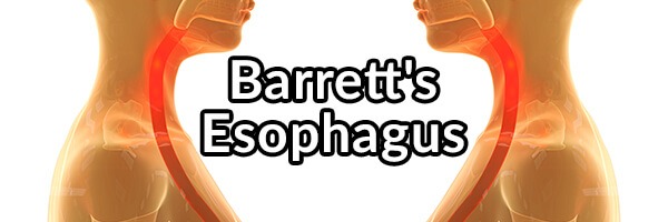
Barrett’s Esophagus, What Is It and What Can Be Done to Reverse It
When someone is suffering from long-term reflux disorders, they become concerned that they might develop esophageal cancer. You get an endoscopy procedure performed because you have reflux and your gastroenterologist tells you that you have Barrett’s esophagus which increases your risk of esophageal cancer. Hearing this troubling news causes you unnecessary stress. Barret’s esophagus diagnosis has risen dramatically in the United States more than 6-fold in 40 years from 0.4 cases per 100000 in 1975 to 2.6 cases per 100000 in 2009. Diagnosis of reflux is also on the rise within our modern world. What is causing the increase in cases, and what are your chances of developing esophageal cancer from Barret’s esophagus?1
What Is and Are the Causes of Barret’s Esophagus?
Barret’s esophagus is named after surgeon Norman Barrett. However, Dr. Philip Rowland Allison described the condition accurately first in 1953. “In 1953, Allison and Johnstone described 7 patients who presented reflux esophagitis involving an “oesophagus lined with gastric mucous membrane” and refuted Barrett’s contention that the tubular, intra-thoracic, columnar-lined viscus was the stomach. Allison and Johnstone’s arguments were eventually accepted by Barrett in a report published in 1957, which suggested that the condition should be called lower oesophagus lined by columnar epithelium.” Allison should have gotten credit and had the disease named after him but alas he did not. Barrett’s esophagus is an abnormal change in the cells of the esophagus. Long-term GERD, LERD, bile reflux, or vomiting may cause the stratified squamous epithelium cells to be replaced by simple columnar epithelium with goblet cells. Bile reflux appears to be the primary cause of Barret’s esophagus and maybe the quickest way out of all the reflux types to develop the condition. Finally, some cases of Barrett’s esophagus are asymptomatic as well.2 3 4
The development of the condition occurs when esophageal cells are exposed to over time pepsin, bile acids (especially bile), or stomach chyme and chronic cellular degradation occurs from inflammation. Instead of the damaged cells dying and being renewed the body attempts to replace the normal esophageal cells (squamous cell epithelium) with cells that secrete more mucus (an increase in goblet cells is seen, they produce mucus) and bicarbonate, cells that closely resemble stomach and duodenal lining cells (columnar epithelium) to protect the esophagus from further damage. It is unknown how the cellular change occurs within the esophagus, however, there are two accepted reported theories. The first theory is that the cellular changes appear to migrate upwards from the gastroesophageal junction (where the lower esophageal sphincter is located), meaning columnar epithelium cells migrate from the LES changing the cellular structure of the esophagus from the bottom, up. The second theory is that are changes in the commitment of pluripotent stem cells lining the esophagus that renew the esophageal tissue from squamous cells to columnar epithelium cells from continuous exposure to reflux. Even though the body is trying to adapt to protect itself from further harm, it comes at a cost because those cells are not supposed to line the esophagus. Bile acids were shown to be able to induce esophageal columnar epithelium differentiation, and the bile acid deoxycholic acid is the most potent in inducing mucosal injury. “Extracted results from in vivo and in vitro studies demonstrate that bile acids are responsible for the increase of reactive oxygen species (ROS) leading to oxidative DNA damage and activating the NF-κB pathway for cell death. The impact of oxidative DNA damage is more severe with bile acids because there is a concomitant decrease in MnSOD expression, which is a scavenger for ROS. Bile acid also induces cytokine-mediated cell damage and may stimulate the expression of COX2, BMP4 and MUC2 genes for proliferation of intestinal metaplasia.“5 6 7 8 9
This change in the cells of the esophagus increases the risk of esophageal cancer formation. The risk, however, is not as much as initially believed at .5%, however, the annual risk is closer to .12% depending on the severity of one’s disease. However, esophageal adenocarcinoma has a high mortality rate. The increased risk of cancer develops from chronic inflammation damaging esophageal cells from increased reflux, changing their differentiation, and leading to inappropriate cellular mutation and survival (cancer). Certain risk factors seem to increase your chance of getting cancer if you have Barrett’s. One such risk factor is consuming alcohol if you have a polymorphism that leads to decreased production of the detoxification enzyme aldehyde dehydrogenase such as an ALDH2 polymorphism. When you consume alcohol if your face flushes, you more than likely have an ALDH2 polymorphism. If you have this polymorphism and are refluxing, it might be best to cease all alcohol consumption until you get your reflux under control. Finally, it appears that is it the reduction of estrogen production (which estrogen decreases cellular growth, reduces differentiation [squamous to columnar cells], and increases cancer cell apoptosis) from consuming alcohol with an ALDH2 polymorphism because alcohol is more harmful to the liver because of impaired detoxification. Therefore, the ALDH2 polymorphism may increase the cancer risk in women only, since men only produce low amounts of estrogen anyway.10 11 12 13 14 15 16
The tissue change in Barrett’s esophagus will reverse itself usually after the causes of the disease are remedied, and the risk of cancer then becomes greatly reduced over time. Barrett’s esophagus remission might occur from stem cell differentiation returning to normal renewing the esophageal tissue with squamous epithelium.17 18 19
Signs of symptoms of Barrett’s esophagus include frequent and long-term GERD/LERD/bile reflux, dysphagia, vomiting of blood, pain under the breastbone, esophageal pain, esophageal burning, and weight loss from eating reduction. If the cause of your Barrett’s esophagus is silent reflux, it may be asymptomatic. Barrett’s esophagus is more common in overweight people and affects men more frequently than women (estrogen production may protect against developing Barrett’s esophagus).20 21
Diagnosis of Barrett’s esophagus is usually made from an endoscopy. Biopsies from the endoscopy are examined under a microscope, and changes of esophageal tissue (inflammation, goblet cells, columnar epithelium tissue) are seen in people suffering from the disease. “Barrett’s esophagus (BE) presents on endoscopy as characteristic salmon-pink colored extensions (or “tongues”) of mucosa that grow into the esophagus above the esophageal gastric junction (EGJ). For screening and surveillance, four quadrant biopsies are taken along every 2 cm of the BE type mucosa and submitted to pathology in separate containers. While this approach samples only a small fraction of the affected lining, it allows the opportunity to recognize dysplasia and focus subsequent biopsies on the appropriate area if dysplasia is identified.” I recommend that you get a transnasal esophagoscopy instead of an endoscopy. It appears that this test is safer, without the risk of the use of anesthesia in routine endoscopies. Someone with Barrett’s esophagus is recommended to get a transnasal esophagoscopy every three years after remission of the disease to make sure the condition remains in remission.22 23 24 25
Barrett’s Esophagus Protocol
Follow GERD, LERD, or bile reflux protocols to remedy underlying condition to reverse Barrett’s esophagus.
Add any supplement below to see if they help if they are not in any of the protocols mentioned above:
- Pure Encapsulations zinc carnosine – take one capsule with a meal, twice daily.
- DGL licorice chewable – follow supplement recommendations on the bottle (take thirty minutes before a meal; chew very well and mix with saliva for effectiveness).
- Georges Always Active aloe vera – follow bottle recommendations.
- Slippery elm lozenges– follow tin recommendations.
- Organic black raspberry powder – consume one teaspoon with a glass of filtered water mixed well, once daily.
- Magnesium glycinate – two to six capsules, taken at bedtime.
- Drink and gargle with two to four ounces of naturally, alkaline water (Evamor and Mountain Valley Spring Water are recommended brands) two hours after a meal to deactivate pepsin in the larynx (from stomach contents reflux) that might cause irritation. In addition, drink and gargle with 1/4 cup of naturally alkaline water before bed.
- Follow D-limonene protocol if no relief in ten days.
Zinc carnosine will help relieve stomach inflammation and dysbiosis and improve the integrity of the LES.
DGL, aloe vera, and slippery elm coat the esophagus and help prevent further reflux inflammation.
Black raspberry powder has high free radical scavenging polyphenol content and seems to reverse Barrett’s esophagus in studies.26 27
D-limonene protects and coats the stomach/esophagus and relieves stomach dysbiosis.
- ↩
- Beers, Mark. The Merck Manual, Merck Research Laboratories, 2006. ↩
- Tamparo, Carol, Lewis, Marcia. Diseases of the Human Body, F. A. Davis Company, Feb. 11, 2011. ↩
- https://www.ncbi.nlm.nih.gov/pmc/articles/PMC4391409/ ↩
- Beers, Mark. The Merck Manual, Merck Research Laboratories, 2006. ↩
- Tamparo, Carol, Lewis, Marcia. Diseases of the Human Body, F. A. Davis Company, Feb. 11, 2011. ↩
- http://www.ncbi.nlm.nih.gov/pmc/articles/PMC3265012/ ↩
- https://www.ncbi.nlm.nih.gov/pmc/articles/PMC5407481/ ↩
- https://www.ncbi.nlm.nih.gov/pmc/articles/PMC4391409/ ↩
- Beers, Mark. The Merck Manual, Merck Research Laboratories, 2006. ↩
- Tamparo, Carol, Lewis, Marcia. Diseases of the Human Body, F. A. Davis Company, Feb. 11, 2011. ↩
- http://www.ncbi.nlm.nih.gov/pmc/articles/PMC3265012/ ↩
- http://www.gastrojournal.org/article/S0016-5085%2808%2902180-X/abstract/ ↩
- http://www.ncbi.nlm.nih.gov/pubmed/16103445 ↩
- http://www.ncbi.nlm.nih.gov/pubmed/22320964 ↩
- http://www.nejm.org/doi/full/10.1056/NEJMoa1103042 ↩
- Beers, Mark. The Merck Manual, Merck Research Laboratories, 2006. ↩
- Tamparo, Carol, Lewis, Marcia. Diseases of the Human Body, F. A. Davis Company, Feb. 11, 2011. ↩
- https://www.ncbi.nlm.nih.gov/pmc/articles/PMC4391409/ ↩
- Beers, Mark. The Merck Manual, Merck Research Laboratories, 2006. ↩
- Tamparo, Carol, Lewis, Marcia. Diseases of the Human Body, F. A. Davis Company, Feb. 11, 2011. ↩
- Beers, Mark. The Merck Manual, Merck Research Laboratories, 2006. ↩
- Tamparo, Carol, Lewis, Marcia. Diseases of the Human Body, F. A. Davis Company, Feb. 11, 2011. ↩
- http://transnasalesophagoscopy.com/ ↩
- https://www.ncbi.nlm.nih.gov/pmc/articles/PMC3418534/ ↩
- http://www.ncbi.nlm.nih.gov/pmc/articles/PMC3079338/ ↩
- http://www.ncbi.nlm.nih.gov/pmc/articles/PMC3047236/ ↩


I just diagnosed with Barrett’s esophagus without dysplasia. What should I do to correct it?
I just bought the book and started to study it. Lot to “digest”. Incredible and very unique work.
I was just diagnosed with hiatal hernia, two duodenum ulcers, Gerd and a starting Barrett’s esophagus… It is a little overwhelming.
My doctor suggested to take daily 20mg Omeprazole “until the rest of my life” and of course watch my diet. I have thought about the same issues regarding the Omeprazole as you are mentioning in your book. The question is, what should I do having all these issues. I am a little worried to stop the PPI although at the moment I had to change to Famotidine 20 mg 2x daily because my tongue started to get numb but when this symptom will go away, my doctor told me to start on Nexium. Should I combine my medicine with some of the things you recommend and if yes, what should those be and how much?
I would very much appreciate to hear your suggestion.
Hi, I was just diagnosed with Barrett’s, and I have to say that this is the most informative article I have read on the subject. I am trying to determine what caused this in the first place; I never had a problem until after menopause. I take HRT and notice a big difference in my symptoms as my hormones fluctuate. Thank you in advance for any advice/information!
Did most of these..my barretts is now gone according to my endoscopy..Please dont forget that prayer is the number one healing factor..then use these supplements..I used zinc carnosine twice daily, Dgl powder twice daily, magnesium glycinate at bed time, aloe vera capsules, l glutamine 5 grams twice daily and drank ginger tea every morning..no wine, coffee, onions, spicy food except on rare occasion..
I just was diagnosed with Barrett’s and hiatal hernia, I’m really worried. I hope I can reverse one if not both of thee conditions.
Thanks for sharing
Karina
Hi Linda,
How long were you on the protocol you mentioned?
Thanks
Adam
Wow. This is great advice. Thanks. I will do these.